by Dr PARVEZ SHEIK
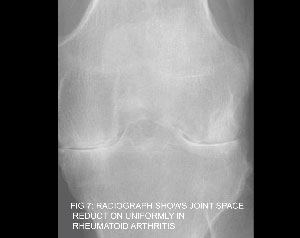
Chronic joint and muscular pain can be distressing and needs attention and care as early diagnosis and treatment has more chances of recovery . Rheumatology encompasses a wide range of medical conditions which cause joint inflammation .Rheumatological disorders may reveal mild to severe changes in the joints on radiological investigations depending upon the course and severity in individualcases . Radiological investigations like Xrays , ultrasound , CT scan , MRI are helpful to detect and also play a role in follow up of patients who suffer form Rheumatological disorders. Xray investigation is very useful modality to screen and follow up patients in Rheumatic disorders.Soft tissue swelling , bone erosions and loss of normal joint space are some features that can be appreciated on xrays. Diagnostic xrays involve the use of radiation and xrays are avoided during pregnancy . Notify your doctor if you are pregnant . Bone densitometry tests also use xray radiation to evaluate the bone loss in long standing rheumatic patients. CT scan or Computed tomography scan is useful to scan the joint and spine and produce high resolution images . High resolution CT scan of chest called HRCT Thorax is helpful in patients who develop chest complications from long standing rheumatological disorders . 3D CT scan is helpful as preoperative investigation for those patients needing surgery. Ultrasound investigation uses high ultrasonic frequency probes to scan and there is no risk of radiation exposure. It is useful for imaging the joints to detect soft tissue inflammation and fluid collection in swollen joints. Ultrasound guided aspiration of inflammed joint fluid can be done . Ultrasound guided steroid injections into joint space is done to reduce inflammation . Ultrasound screening of abdomen can detect pre-existing diseases in the liver and kidney , pelvic infections in females , free fluid collection in abdomen and pelvis. Magnetic resonance imaging (MRI ) uses high magnetic fields to generate images of the joints , there is no risk of radiation involved . MRI in rheumatology helps in visualizing the spine and joints better than other imaging modalities as the soft tissue resolution is excellent . Small magnetic coils which direct high magnetic field are placed near inflammed joint and images are reconstructed using a special computer software . However if the patient has artificial metal implants or metallic stents then MRI should be avoided. Radiological aspects in Rheumatological disorders: Rheumatoid arthritis affects the small joints like those in the hand and foot , can involve the large joints like the knee and hip . Xrays may show soft tissue swelling , small joint erosions , loss of joint space ,osteopenia . Ankylosing spondylitis mostly affects the spine . There is loss of normal movement in spine as the disease progresses. Xrays of spine may show calcification in ligaments around the vertebrae but disc space is preserved. Fusion of the posterior elements in spine may occur. Psoariatic arthritis is a skin disorder that also affects the joints like those in the hand and foot . Xrays may show erosive bone lesions and soft tissue swelling. Gouty arthritis occurs when serum uric acid levels are high and calcific trophi get deposited in the small joints of hand and foot. Xray of hand and foot may show the calcified gouty trophy at small joints, bony erosions and soft tissue swelling. Sacroiliitis is inflammation of sacroiliac joint located in the lower back region . Sacroiliitis can cause pain in lower back and can extend down one or both legs. Activities like prolonged standing or climbing steps can worsen the pain.. Xrays would show sclerotic inflammatory changes at sacroiliac joints. Systemic lupus erythematosis (SLE ) is a chronic systemic disease which can affect the joints . Patients can have sun sensitivity rashes, hair loss , swelling or tenderness of the small joints of the hands, feet,knees, and wrists, ulcers in mouth , fluid collection in chest and cardiac regions . Hand xrays may show soft tissue swelling of the involved joints, periarticular osteoporosis, and normal joint spaces. Scleroderma, also known as systemic sclerosis , is a multi-system autoimmune connective tissue disorder. Xray of Hands may show joint space narrowing , erosions, periarticular osteoporosis , soft tissue calcification. Juvenile rheumatoid arthritis, is the most common chronic arthritic disease of childhood . Joint pains may involve large and small joints of more than 6 weeks duration. X-ray of hand may show soft tissue swelling, osteopenia, loss of joint space, erosions and joint subluxation. Cervical spine xrays may show subluxation and erosions at 1st and 2nd cervical vertebrae, fusion of the posterior elements of cervical vertebrae. Osteoarthritis is degenerative disorder of joints . Sometimes in long standing cases of inflammatory joint disorders like Rheumatoid arthritis may progress and cause degenerative changes at joints . Xrays may show osteophyte formation, joint space narrowing, subchondral sclerosis and cysts.
Figure.1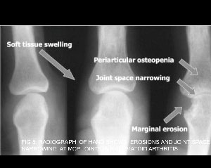
Figure.2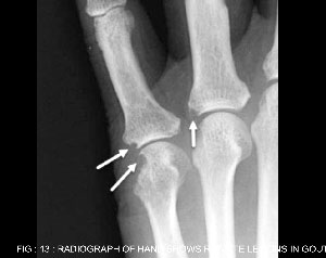
Figure.3